If you have severe upper jaw bone loss and want fixed teeth without grafting or a sinus lift, pterygoid implants offer a reliable alternative by anchoring into the dense pterygoid region behind the maxilla. These implants let you avoid lengthy grafting procedures and often shorten treatment time while still providing stable support for prosthetic teeth — making them one of the more advanced full mouth dental implants in Woodbury, MN.
Pterygoid implants work by engaging strong posterior bone to secure implants when the usual back-of-jaw bone is insufficient, so you can skip sinus augmentation in many cases. The article will walk through the anatomy and surgical approach, compare pterygoid implants with sinus lifts, explain who qualifies, and summarize long-term outcomes and recent innovations so you can weigh this option confidently.
Anatomy and Mechanism
You will see that the pterygoid region provides dense cortical bone, a specific angulation for implant insertion, and direct posterior anchorage that bypasses the maxillary sinus. These features determine surgical access, prosthetic emergence, and long-term load distribution.
Pterygoid Bone Structure
The pterygoid region comprises the maxillary tuberosity, the pyramidal process of the palatine bone, and the pterygoid process of the sphenoid. You rely on the dense cortico-cancellous junction near the pterygoid plates for primary stability.
Bone quality here is typically denser than the posterior maxillary alveolus, reducing the need for grafting.
Key anatomical landmarks to identify on CBCT:
- Maxillary tuberosity for initial entry point.
- Pterygoid plates (medial and lateral) for anchorage depth.
- Greater palatine foramen and neurovascular bundles to avoid.
You must assess the posterior superior alveolar artery trajectory and the pterygomaxillary fissure to avoid hemorrhage or sinus perforation. Precise angulation and depth planning optimize engagement with cortical bone while protecting adjacent anatomy.
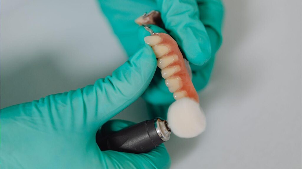
Implant Placement Technique
You position the implant at a 45°–60° posterior-medial angulation relative to the occlusal plane. This trajectory allows the implant to traverse the tuberosity and engage the pterygoid plate without entering the sinus cavity.
Use a staged drilling protocol with decreasing diameters to preserve bone and increase insertion torque.
Procedural steps you follow:
- Mark soft-tissue incision posterior to the tuberosity.
- Create a pilot osteotomy aimed at the pterygoid plate under CBCT guidance.
- Sequentially widen the osteotomy and place a longer implant (usually 13–20 mm) to engage cortical bone.
- Verify position radiographically and manage soft-tissue emergence for prosthetic access.
You may choose an angled abutment or prosthetic cantilever depending on arch spread. Atraumatic technique and real-time imaging reduce risks of vascular or sinus complications.
Biomechanical Advantages
Pterygoid implants convert posterior vertical deficiency into a posteriorly anchored fulcrum, increasing anteroposterior (AP) spread of the prosthetic base. You gain 8–12 mm or more of AP extension compared with standard posterior implant positions, improving load distribution.
Direct engagement with dense cortical bone increases primary stability and allows immediate or early loading in many cases.
Mechanical benefits you achieve:
- Reduced cantilever forces on anterior implants.
- Lower need for sinus lifts or bone grafts.
- Enhanced resistance to axial and lateral masticatory loads.
Monitor torque values and peri-implant bone remodeling with periodic radiographs to confirm sustained biomechanical performance.
Comparison With Sinus Lift Procedures
Pterygoid implants let you avoid grafting in many posterior maxilla cases by anchoring into the pterygoid plate, while sinus lifts build vertical bone height under the maxillary sinus to support standard implants. Each option changes surgical access, healing time, and long-term maintenance in specific ways.
Indications for Each Approach
Pterygoid implants suit you when the posterior maxilla has severe vertical bone loss but adequate pterygoid plate anatomy and when you want to avoid grafting. They work well for patients needing fixed posterior support immediately or within a shortened treatment timeline. You need good mouth opening and an experienced surgeon because the angle and length of these implants demand precise placement.
Sinus lifts suit you when you prefer standard implant positions and crowns on shorter implants, or when the sinus floor is low but lateral bone quality allows grafting. You may choose a sinus lift if anatomy or access precludes safe pterygoid implant placement, or when long-term familiarity with standard implants is a priority for your surgeon.
Recovery Times and Outcomes
Pterygoid implants typically shorten overall treatment time because you often skip grafting and wait periods. Immediate or early loading is possible in many cases, which lets you receive fixed prostheses sooner. Expect postoperative soreness in deeper tissues and a learning-curve for hygiene, but studies report high survival rates comparable to conventional posterior implants when placed correctly.
Sinus lift procedures add about 4–8 months to treatment because graft maturation precedes implant placement. Your recovery includes sinus-specific risks such as membrane perforation and longer soft-tissue healing. When successful, sinus lifts permit standard implant placement and familiar prosthetic workflows, with predictable long-term function if graft integration proceeds without complications.
Limitations and Contraindications
Pterygoid implants are technique-sensitive. You should avoid them if you have unfavorable pterygoid anatomy, limited mouth opening, or active infection in the posterior maxilla. Hygiene access can be more challenging for some prosthesis designs, and rehabilitation requires careful occlusal planning to avoid biomechanical overload.
Sinus lifts carry surgical risks like membrane tears, graft infection, and delayed implant placement due to graft failure. You may not be a candidate if you have chronic sinus disease, heavy smoking, or systemic conditions that impair bone healing. Both approaches require individualized radiographic evaluation (CBCT) and treatment planning by a clinician experienced in the chosen technique.
Patient Suitability and Evaluation
You need a focused clinical exam, targeted imaging, and assessment of local and systemic factors to determine if pterygoid implants suit your case. The evaluation identifies anatomical landmarks, bone quality, and risks that affect planning and long-term success.
Clinical Assessment Process
Start with a thorough dental and medical history. Record prior sinus disease, smoking, bisphosphonate use, radiation, and uncontrolled diabetes because these affect healing and infection risk.
Perform an intraoral exam to check ridge form, residual tuberosity height, mucosal health, and keratinized tissue. Palpate the tuberosity and pterygoid region for undercuts, soft-tissue thickness, and proximity to the greater palatine artery.
Assess occlusion, parafunction (bruxism), and opposing dentition to determine loading demands. Measure interarch space for prosthetic clearance and plan whether a fixed or removable restoration will be supported.
Discuss patient expectations, treatment timeline, and tolerance for local versus more invasive alternatives. Obtain informed consent after explaining risks specific to the pterygoid area, such as neurovascular proximity and access limitations.
Radiographic Criteria
Obtain a CBCT as the primary imaging modality for 3D assessment of the pterygoid pillar, maxillary tuberosity, and sinus boundaries. Use thin slices (0.2–0.3 mm) and multiplanar reconstructions to visualize cortical engagement points.
Evaluate bone height, width, and cortical density at the pterygoid plate and tuberosity. Look for sufficient posterior-lateral bone to allow a 13–20 mm implant trajectory toward the pterygoid pillar without penetrating the sinus or infratemporal fossa.
Identify critical landmarks: pterygoid fissure, greater palatine canal, maxillary sinus floor, and zygomatic buttress. Measure distances from the planned apex to the greater palatine artery and pterygomaxillary fissure to reduce vascular or soft-tissue injury.
Use surgical guides or virtual planning when anatomy is complex. If CBCT shows insufficient bone or unfavorable angulation, consider alternative strategies (sinus lift, short implants, or zygomatic solutions).
Factors Influencing Success
Bone quality at the pterygoid region strongly affects primary stability; dense cortical engagement yields better immediate fixation. Aim for bicortical or multi-cortical anchorage when possible.
Patient systemic health and habits matter: smoking and poorly controlled systemic disease increase failure risk. Manage modifiable risks preoperatively and ensure good oral hygiene and periodontal control.
Surgical skill and accurate trajectory control impact complication rates. Proper training in the pterygoid approach and use of guided or pilot-drill protocols reduce implant malposition.
Prosthetic planning—load distribution, splinting implants, and occlusal scheme—affects long-term survival. Plan for rigid splinting to minimize lever forces on posterior implants when opposing dentition produces high occlusal loads.
Long-Term Outcomes and Innovations
Pterygoid implants routinely provide posterior support without sinus lifts, showing predictable survival and functional stability. Complication patterns tend to be limited and manageable, while new device designs and guided techniques are reducing surgical variability.
Success Rates and Stability
Clinical studies report high survival rates for pterygoid implants, often comparable to implants placed in other maxillary regions over one to nine years. You can expect survival rates commonly reported in the 90%+ range when implants engage the dense cortical bone of the pterygoid plates and when clinicians follow standardized placement protocols.
Primary stability is typically greater than for implants placed in atrophic posterior maxillae because of bicortical engagement. This stability shortens or eliminates the need for delayed loading in many full-arch rehabilitations. Prosthetic maintenance is similar to other full-arch restorations; control of occlusal forces and regular follow-up enhance long-term prognosis.
Complication Management
Early complications include transient postoperative pain, swelling, or minor paresthesia; these usually resolve within weeks. Infection and implant failure are uncommon but require prompt diagnosis—antibiotics, drainage, or implant removal when clinical signs persist.
Technical complications include prosthetic screw loosening and wear on angled abutments. You can manage these with routine occlusal checks, torque verification, and component replacement when indicated. Imaging (CBCT) helps detect maladaptive angulation or bone loss early, guiding corrective measures such as prosthetic adjustment or, rarely, implant removal.
Emerging Techniques in Implantology
Guided surgery and CAD/CAM surgical guides are increasing accuracy for pterygoid trajectories and reducing operative time. You can use dynamic navigation or stereolithographic guides to plan angulation that optimizes pterygoid plate engagement while avoiding adjacent anatomic structures.
Implant design innovations—longer tapered bodies, surface treatments to enhance osseointegration, and specific angulated prosthetic connections—improve immediate stability and prosthetic fit. Minimally invasive access protocols and local hemostatic strategies reduce morbidity. Researchers are also exploring digital workflow integration from planning to prosthesis delivery to shorten treatment timelines and improve reproducibility.