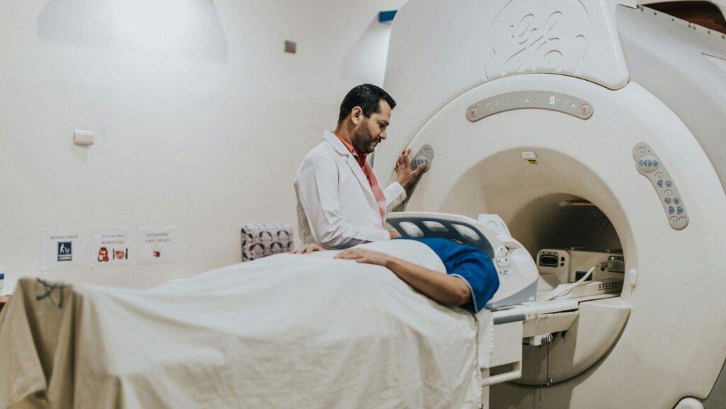
If you're planning dental implants, you want precision—and results you can count on. A CT scan maps your jaw in 3D, showing bone volume, density, nerves, and sinuses. That way, your dentist can place implants more safely and accurately than with old-school 2D X-rays.
Let's talk about how CT imaging guides surgical planning, cuts down on risks like nerve injury or sinus trouble, and works with digital guides and implant tech for better results. You'll get a clearer sense of why modern implant care leans on cone-beam CT, and what that means for your treatment, safety, and long-term success—and if you're ready to explore your options, connecting with experienced dentists in New Market VA is a great place to start.
Role of CT Imaging in Dental Surgery
CT imaging gives you a precise 3D look at bone volume, nerve and sinus locations, and any local issues. This info directly guides where to place implants, whether you need grafting, and how to avoid problems during surgery.
Accuracy in Assessing Jawbone Structure
Cone beam CT (CBCT) shows voxel-level detail in your upper and lower jaw. You can measure bone height, width, and density at possible implant sites.
Cross-sectional and axial views let you check ridge dimensions to the millimeter and see if the bone's thick enough for stability.
Spotting thin bone early means you can plan grafts or pick shorter or narrower implants—no guessing on the day of surgery.
You get a real bone map, which cuts down the risk of implant failure from poor integration.
Identification of Anatomical Landmarks
CT scans help you find the inferior alveolar nerve canal, mental foramen, maxillary sinus floor, and nasal cavity in 3D.
You can measure how close your planned implant is to these structures and set safety margins—usually 1–2 mm from nerves and sinuses.
You can export DICOM data into planning software to simulate implant angle and depth, while seeing nearby nerves and blood vessels.
Surgical guides made from this data turn the plan into precise drilling paths in your mouth, so there are fewer surprises during surgery.
Visualization of Pathology or Irregularities
CT imaging uncovers cysts, leftover roots, periapical lesions, and sinus disease that 2D X-rays often miss.
You can spot infections or bone defects that need fixing before placing implants, which lowers the risk of problems after surgery.
You might also see oddities like accessory canals, thin buccal plates, or bone loss that affect your implant choice and surgery approach.
Catching these early lets you plan extra steps, change implant spots, or tweak the prosthetic plan to protect your long-term results.
Advantages Over Traditional X-Rays
CT imaging gives you a real 3D view of bone, nerves, and sinuses.
That means fewer surprises during surgery and more precise, custom implant placement—often saving time in the chair.
Three-Dimensional Visualization
CT scans (including cone-beam CT) produce 3D images, so you see your jaw from all angles: axial, coronal, and sagittal.
You can check bone height, width, and density right where you need to place the implant, not just guess from a flat X-ray.
You see exactly how close you are to the nerve, foramen, or sinus, measured in millimeters.
Knowing those distances helps you pick the right implant length and angle to avoid nerve or sinus trouble.
You can digitally slice and rotate the image, which makes it easier to spot uneven bone loss, tilted roots, or old hardware.
These details matter when deciding on grafting, short implants, or angled components.
Minimized Risk of Complications
CT-based planning cuts the risk of nerve damage, sinus perforation, and implant misplacement by giving you clear spatial data.
Measuring canal-to-implant clearance in millimeters lets you set real safety margins, not just guess from a 2D film.
CT reveals bone defects or thin plates that look fine on regular X-rays.
If you spot these before surgery, you can change the plan—pick a different implant, add grafting, or stage the case—to avoid complications.
CT also shows nearby problems like cysts or impacted teeth that could mess up healing.
Fixing these first means fewer infections and less chance you'll need more surgery later.
Enhanced Surgical Planning
You can import CT data into planning software, place virtual implants, and check angles.
You can try out different sizes and positions, and instantly see how each one fits the bone and nearby teeth.
Surgical guides based on CT plans translate those virtual positions into real-life accuracy, often within a millimeter.
That kind of precision saves time and means fewer changes during the procedure.
Planning ahead with these details helps you, your surgeon, and the lab stay on the same page.
You can sort out grafts, guide types, and prosthetic parts before the appointment, which streamlines everything.
Integrating Digital Data With Implant Technology
You can combine 3D imaging, intraoral scans, and prosthetic files to make implant placement more accurate and predictable.
This integration makes planning smoother and helps the final restoration fit better—both functionally and esthetically.
Use in Computer-Guided Surgery
CBCT volumes can be matched with your intraoral scans to build a patient-specific plan showing bone, nerves, and where the teeth will go.
You can place virtual implants in 3D, check how close they are to nerves or sinuses, and pick the right size and length for your anatomy.
Guided surgery systems turn that plan into drill sequences and sleeve positions.
Static guides lock in the drill angle and depth; dynamic navigation shows your drill tip on a monitor as you work.
Both methods help keep you on the planned path, which is especially helpful in tricky or full-arch cases.
It's smart to double-check registration accuracy and make sure the plan matches the guide before starting.
Export STL files for guide printing and keep a checklist: patient orientation, guide fit, and drill stop lengths, so the digital plan becomes reality.
Collaboration With CAD/CAM Systems
You can export CBCT DICOM and intraoral STL files into CAD software to merge the jaw anatomy with the prosthetic design.
This fusion makes sure the implant lines up with the planned crown emergence and bite.
CAD/CAM lets you review and tweak tooth shape, gum contours, and implant angle, then check the bone again.
Clinicians and dental techs can use digital files and cloud models, which speeds up communication and reduces mistakes.
Most workflows let you generate implant libraries, abutment designs, and temporary prosthetics from this merged data.
That means you get faster lab turnaround and temporaries that actually match the final look.
Customization of Surgical Stents
You can design surgical stents from the merged CBCT and prosthetic models to control depth and angle.
Customize guide features—open or closed sleeves, support type, and irrigation holes—to fit your case.
Material matters: rigid resin printed at high resolution keeps sleeves accurate, and reinforced designs don't bend during drilling.
Add markers or windows for checking fit, and make cutouts for tension relief if you're working with toothless arches.
Send a checklist to your lab or in-house printer: implant system, sleeve size, and fit points.
Always check the stent in the mouth before drilling to make sure it fits and matches the digital plan.
Patient Outcomes and Safety Considerations
CT imaging boosts surgical accuracy, lowers risk to nerves and sinuses, and gives you a better idea of the long-term outlook and possible issues before surgery starts.
Reduced Postoperative Issues
CT scans let your dentist map bone volume, density, and nerve and sinus positions in 3D.
That info cuts the risk of nerve injury, sinus perforation, and misplaced implants.
Using CT-based guides or navigation, your implant angle and depth become more predictable.
Predictability means fewer immediate issues like bleeding or numbness, and less need for follow-up surgery.
CBCT also helps spot cysts or leftover roots that could mess up healing.
Fixing these before placing implants lowers the chances of infection or failure after surgery.
Informed Consent and Communication
CT images give you visuals you can look at with your clinician.
Seeing your 3D anatomy helps you understand where implants will go, what's nearby, and if you need grafts.
When risks are measured—like how many millimeters from a nerve—your consent is more informed.
You can weigh options like staged grafting, shorter implants, or different prosthetic plans.
Clinicians can document the plan using CT screenshots and details, which makes things more transparent.
That documentation helps with shared decision-making and keeps misunderstandings from getting in the way.
Long-Term Success Rates
Preoperative CT assessment lets clinicians place implants precisely where bone volume and quality are good.
This careful placement helps avoid biomechanical overload and uneven force distribution—two things that often lead to implant failure down the line.
When CT guides graft planning, clinicians can figure out graft size and placement with much more confidence.
That accuracy makes it a lot easier to get better osseointegration.
Better graft outcomes usually mean implants survive longer, sometimes for years.
I've seen evidence that 3D planning leads to fewer complications and revisions, especially in tricky cases near vital structures.
CT can't promise perfect results every time, but it really does improve the anatomic factors that matter for long-term stability.